

Proximal Femoral Nail Antirotation (PFNA): Technique, Indications & Instrumentation
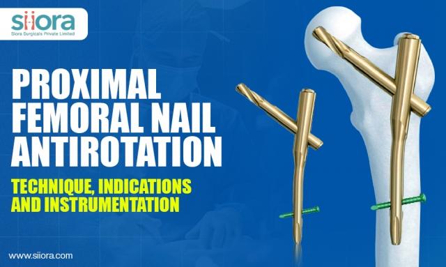
Fractures around the hip, especially those near the upper part of the femur, can be tricky to treat, particularly in older adults whose bones may be weaker. Among the different ways surgeons fix these fractures, the Proximal Femoral Nail Antirotation, or PFNA, stands out because it combines strong fixation with a minimally invasive approach. Let's explore how PFNA works, when it's used, and what surgical tools make the procedure possible.
What Is PFNA?
Simply put, PFNA is a special kind of nail that goes inside the thigh bone (femur) to stabilize fractures near the hip. Unlike traditional nails, PFNA includes an antirotation blade (instead of a regular screw) that locks into the bone, providing extra resistance against twisting forces. This means fractures can heal more securely, which is especially important in unstable breaks where rotation could lead to failure.
When Do Surgeons Prefer PFNA?
PFNA is primarily chosen for fractures that occur in the upper part of the femur, specifically:
- Intertrochanteric fractures: These happen between two bony prominences called the greater and lesser trochanters. In cases where the fracture is unstable or has complicated patterns, PFNA often offers better fixation compared to plates or screws alone.
- Subtrochanteric fractures: These are breaks just below the trochanters. PFNA can support these fractures, especially when intramedullary fixation is preferred to manage the high stress in this area.
- Osteoporotic bone: Older adults often have brittle bones, and the PFNA's blade design compacts the bone as it's inserted, providing superior grip compared to standard screws.
- Certain periprosthetic fractures: Although less common, PFNA may sometimes be used near hip implants to stabilize fractures.
Breaking Down the PFNA Surgical Approach
The success of PFNA largely depends on the surgeon's technique. Here's a simplified outline:
- Planning: Surgeons carefully study X-rays or CT scans to understand the fracture pattern and decide on nail length and diameter.
- Positioning the Patient: The patient lies on their back on a fracture table, often with some traction applied to help align the broken bone parts.
- Reducing the Fracture: Using gentle manipulation and X-ray guidance, the surgeon attempts to bring the fragments into proper position without making a big cut.
- Entry Point & Nail Insertion: A small cut is made near the top of the thigh bone at the greater trochanter. Through this, the surgeon creates an entry to insert the nail into the canal running down the middle of the femur.
- Introducing the PFNA Nail: The nail slides down the hollow center of the bone, extending sufficiently to stabilize the entire fractured area.
- Blade Placement: Instead of a simple screw, a helical (spiral) blade is inserted to lock the nail into the femoral head, compressing the bone and preventing twisting.
- Distal Locking: Screws placed at the far end of the nail stop it from moving inside the bone shaft.
- Closure & Recovery: After everything is confirmed with X-rays, the incisions are closed. Patients are encouraged to start moving as soon as it's safe, which helps avoid complications from prolonged bed rest.
The Instruments Behind PFNA
PFNA requires specific instruments that make the procedure straightforward and precise:
- Guidewires: These thin wires help the surgeon navigate the nail and blade into exact positions.
- Reamers: Used to slightly widen the intramedullary canal to fit the nail.
- Insertion tools: Specialized handles and jigs assist in controlling and placing the nail smoothly.
- The Helical Blade: The defining feature of PFNA, designed to compress bone and prevent rotation, improving stability over traditional screws.
- Locking Screws and Drills: These ensure the nail stays firmly fixed in the bone.
Throughout surgery, continuous X-ray (fluoroscopy) guides each step without the need for a big incision.
Why Choose PFNA?
There are many reasons why PFNA has gained popularity:
- Strong and stable fixation: Especially important in bones weakened by age or severe fracture patterns.
- Less invasive: Smaller cuts mean less tissue damage and faster healing.
- Reduced complications: Lower risk of the implant loosening or “cutting out” compared to some other fixation devices.
- Early mobility: Patients can often bear weight sooner, which is crucial for elderly patients to avoid issues like blood clots or pneumonia.
Final Thoughts
The PFNA technique represents an important leap forward in managing difficult hip fractures, offering a blend of strength, stability, and minimally invasive approach. For orthopedic surgeons, mastering this technique allows them to provide patients with not only reliable fracture fixation but also a faster recovery path. As with any surgical method, success depends on careful planning, patient selection, and surgical precision, all of which come together to improve outcomes in these challenging cases.
If you are looking to become a trusted orthopedic implants supplier in Saudi Arabia, contact Siora Surgicals Pvt. Ltd., a renowned orthopedic manufacturer in India with a global footprint.